Do you know the main components of a joint?

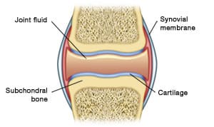
Subchondral bone
The part of the bone located just above the cartilage is called the "subchondral bone". It is "the foundation" of cartilage.
The cartilage
Cartilage is a tissue that forms the lining of the bone ends. It has a pearlescent appearance and its thickness varies from one joint to another. Thus it is the kneecap that has the thickest cartilage: more than 5 millimetres. It is also worth knowing that men's cartilage is generally thicker than that of women.
The role of cartilage is to allow perfect sliding between the bones since the frictional forces are less than that of an ice skater sliding on ice. It is also essential for cushioning and distributing pressure on the bones thanks to its properties of elasticity and resistance.
Cartilage is a living tissue, in perpetual renewal, even among older people: it renews itself completely around every 3 months. Hence, microscopic cartilage fragments constantly break away from the cartilage and end up in the joint cavity (where they are then eliminated by the synovial membrane).
At microscopic level, it is composed of two components:
- On one hand, a matrix consisting of a gel rich in water and molecules called proteoglycans held tightly together in the meshes of the collagen fibre network. Proteoglycans provide the elasticity of the cartilage while the collagen fibres give it resistance to compressive forces.
- On the other hand, cells called chondrocytes, whose role is fundamental because they have the dual ability to renew, but also to destroy the matrix.
The cartilage tissue has no blood vessel and it is not innervated.
The joint fluid
Joint fluid also called synovial fluid is secreted by the synovial membrane. It is present in all joints in small quantities (1 to 2 ml in the knee for example).
Its role is to lubricate the joint. It ensures perfect sliding between the bone ends.
In its normal state it is a clear, transparent fluid with a particular viscosity. The latter is due to the amount of hyaluronic acid present in the joint.
The capsule and the synovial membrane
The capsule and synovial membrane are closely linked since the synovial membrane coats the inside of the capsule, a fibrous covering surrounding the joint.
The synovial membrane is a vascularised and innervated tissue. It is in the form of a smooth transparent membrane covered with small blood vessels. in its normal state it has folds called villi.
Its main role is to secrete the hyaluronic acid of the synovial fluid, a true lubricant of the joint. The synovial membrane also ensures "emptying" of cartilage debris found in the joint cavity. This is thanks to its blood vessels that bring oxygen and nutrients, including glucose, essential for the life of the cartilage.
At microscopic level, the synovial membrane is composed of cells called synoviocytes. It is these cells that perform the functions mentioned above. Lastly, these cells also have the ability to produce enzymes that can destroy the cartilage matrix.


