


Hip: Radiological examinations
X-rays are an essential tool for the diagnosis of osteoarthritis and its medical care.
They are easily interpreted by your doctor. They consist of:
- Narrowing of joint space located in the pressure area of the main knuckle.
- The presence of osteophytes that expand the joint contours.
- Condensation of the subchondral bone (located below the cartilage) in the region of the joint space narrowing.
- Geodes that are inconsistent and established in the condensation (rounded appearance of areas, grey in the condensation).
- At an advanced stage, there is erosion of the subchondral bone located in the pressure zone where the cartilage has disappeared.
If you would like to know more about X-rays and osteoarthritis, click here .
X-rays are used to estimate the deterioration of cartilage and bone caused by osteoarthritis.
However, be aware that there is no real connection between the lesions (described above) observed and clinical signs such as pain or functional impairment.
X-rays of the symptomatic joints are carried out during the first consultation. The frequency for renewing this examination depends on how quickly your osteoarthritis evolves. But it's up to your doctor to determine when they should be done.
The type of X-rays most often requested are images of the pelvis from the front and the so-called Lequesne's false profile view where the patient is standing. The hips may also be X-rayed separately.
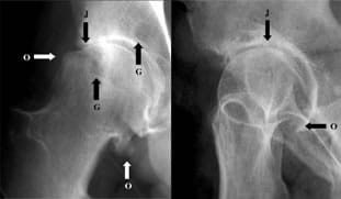
Image of the hip from the front Image of the hip (Lequesne's false profile)
X-rays enable:
- measuring joint space narrowing. This is done at different sites on the hip according to the hip osteoarthritis,
- to identify dysplasia,
- to identify a tendency to coxa valga.
The measurement of joint space narrowing is a good indicator of the level of wear on your cartilage by osteoarthritis.