


Knee: Radiological examinations
X-rays are an essential tool for the diagnosis of osteoarthritis and its medical care.
They are easily interpreted by your doctor. They consist of:
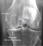
- Narrowing of joint space located in the pressure area of the main knuckle.
- The presence of osteophytes that expand the joint contours.
- Condensation of the subchondral bone (located below the cartilage) in the region of the joint space narrowing.
- Geodes that are inconsistent and established in the condensation (rounded appearance of areas, grey in the condensation).
- At an late stage, there is erosion of the subchondral bone located in the pressure zone where the cartilage has disappeared.
If you would like to know more about X-rays and osteoarthritis, click here .
X-rays are used to estimate the deterioration of cartilage and bone caused by osteoarthritis.
However, be aware that there is no real connection between the lesions (described above) observed and clinical signs such as pain or functional impairment.
X-rays of the symptomatic joints are carried out during the first consultation. The frequency for renewing this examination depends on how quickly your osteoarthritis evolves. But it's up to your doctor to determine when they should be done.
The types of X-rays carried out most often are:
- standing,
- knee fully extended,
- front shot,
- Lyon Schuss radiographic view of the knee,
- generally, both knees are x-rayed separately.
However, these characteristics are the most common, depending on your case, your radiologist may do other views that require other positions.
Other images that are commonly used are in profile and the tibiofemoral view.
Frontal knee X-ray
Measurement of joint space narrowing is a good indicator of the level of wear on your cartilage by osteoarthritis. This is done where the narrowing is the greatest between the two bones in the tibiofemoral compartment or the centre of the tibial plateau.